Introduction
Around 2016, I became interested in microscopy again. I have an educational-level optical microscope, but wanted to take photos and videos with it, and particularly, the 100x objective lens (designed to be used with immersion oil).
Camera/Smartphone Attachment
This is certainly not the best solution to the issue, but it’s nearly universal. The most difficult part of imagine through the eyepiece is holding the camera normal to the eyepiece. I used rubber bands to hold the iPhone against this printed part, which allowed me to shift it around precisely, aligning the exit pupil with the camera lens.
Design:
The below model is press-fit onto the microscope eyepiece:

Dark-Field Microscopy
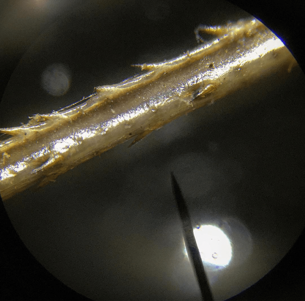
I experimented with foreground illumination. This is useful for solid objects. In most cases, a flashlight or shop lamp provided this oblique illumination.
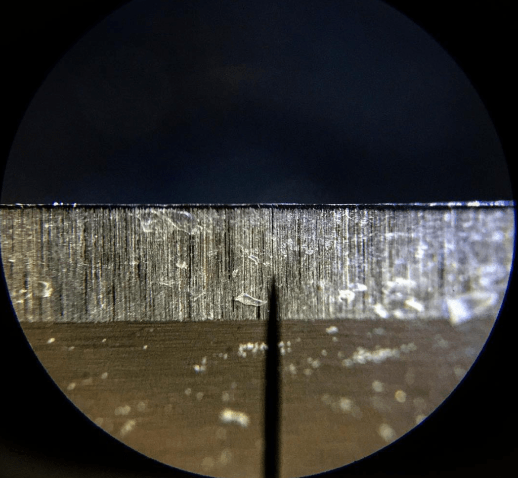
Improper Tempering of Razor Blade Edges:
Here, you can see why inexpensive razor blades may not stay sharp very long; it appears that the blade lost its heat-treatment during the sharpening stage of manufacturing (note the blue oxide):
Reaction of CuSO4, iron filings, and water:
CuSO4 and iron react in a way that deposits elemental copper on iron particles. Here, you can see that reaction in a time-lapse video:
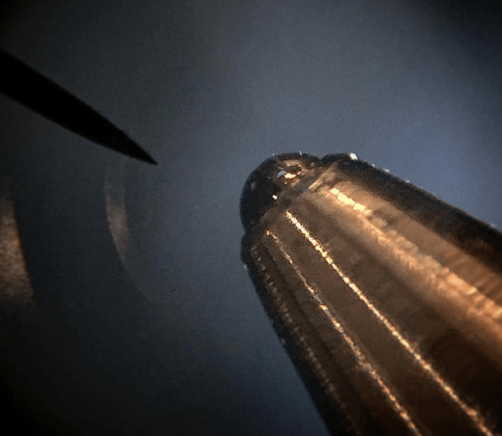
Ballpoint Pen
Out of curiosity, I put a ballpoint pen under the microscope. These pens often use solid carbide spheres for the balls.
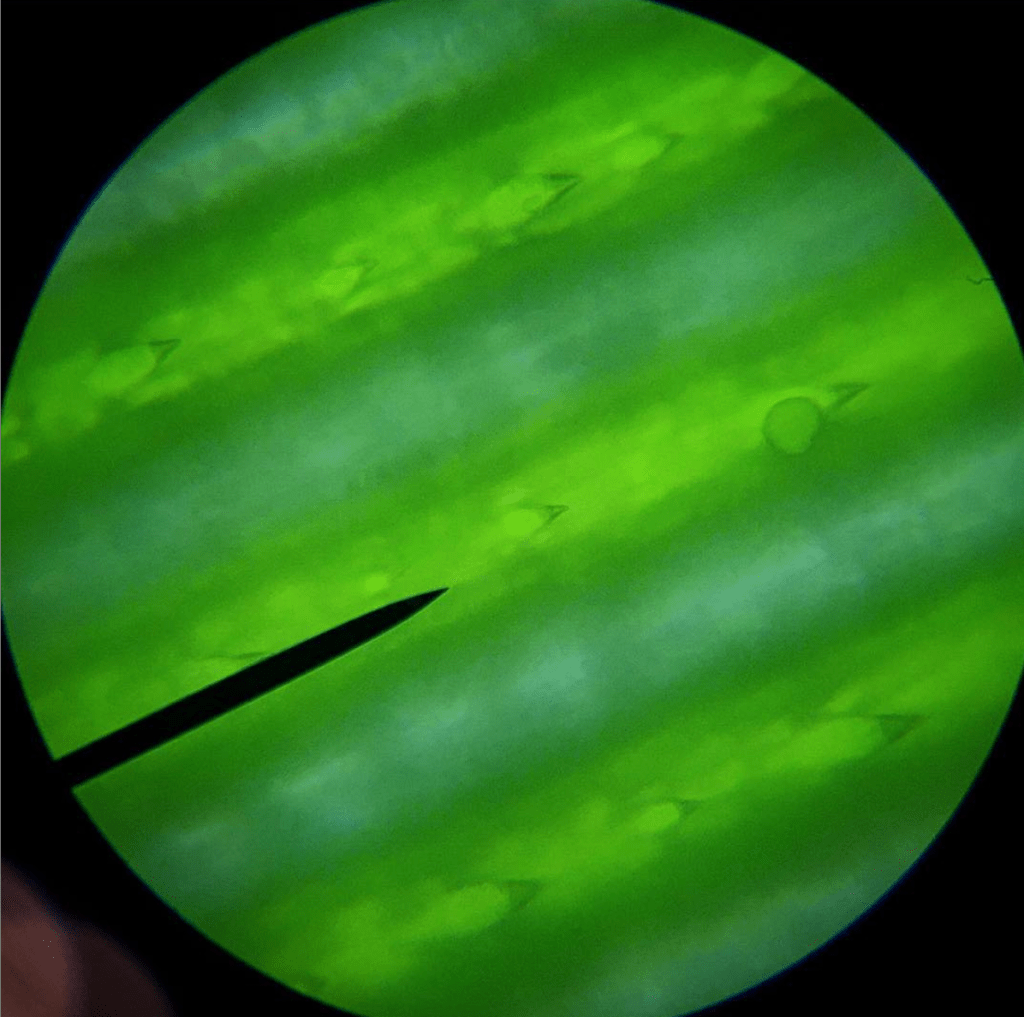
Insects & Plants
Naturally, I placed some insects and plant material under the lens:
Bright-Field Microscopy
As this microscope is intended to be used as a “traditional” bright-field microscope, I spent plenty of time preparing slides and testing the capabilities of the camera system.
Brownian Motion
Brownian motion was visible in the fat globules of homogenized whole milk:
White blood cells (basophils)
I had minor cut and decided to see what my own blood looked like under 100x magnification, using immersion oil:
